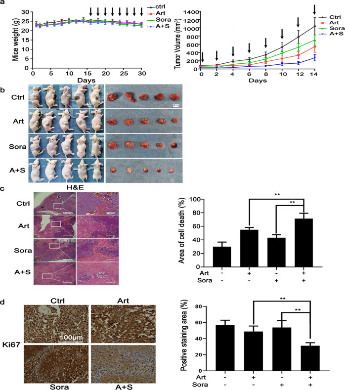
Fig. 2. The combined treatment inhibited xenograft tumors in vivo through extensive cell death.
a Once the size of the inoculated xenograft nodules reached 80–100 mm3, the mice were randomly divided into four groups (n = 5 per group) and treated by gavage with Art (30 mg/kg body weight), Sora (20 mg/kg), the combination of Art and Sora or PBS (control) every other day. The weight of the mice (left panel) and tumor growth (right panel) were monitored regularly. The arrows indicate drug treatment. b The mice were sacrificed, and the tumors were resected for subsequent experiments. c Xenograft tumor sections were stained with H&E. The area of cell death was indicated by smeared cell morphology and faint nuclear staining. The area of cell death is presented in the right panel. d Tumor cell proliferation status was analyzed by IHC staining for Ki-67. **P < 0.01.

