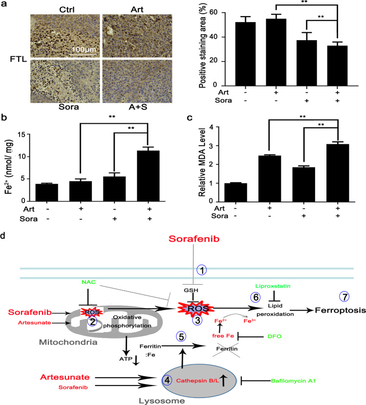
Fig. 6. The combined treatments caused consistent changes in FTL degradation and MDA production in xenograft nodules in vivo.
a Xenograft nodules were collected as shown in Fig. 2b. FTL protein expression was determined by IHC staining as shown in Fig. 2d. The contents of free Fe2+ ions (b) and MDA (c) in tumor nodules were quantified with respective biochemical assay kits. **P < 0.01. d The cartoon describes the molecular mechanisms through which the combined treatment led to ferroptosis. Sorafenib directly inhibits ① GSH synthesis and ② mitochondrial functions, which contributes to ③ extensive oxidative stress. Artesunate primarily ④ promotes lysosomal activation and then synergizes with sorafenib to activate cathepsin B/L activities followed by ⑤ ferritin degradation. The convergence of sorafenib-induced oxidative stress and artesunate-mediated ferritinophagy causes ⑥ lipid peroxidation and eventually ⑦ ferroptosis. DFO, deferoxamine mesylate; GSH glutathione, NAC N-acetyl cysteine; ROS reactive oxygen species. The red color indicates ferroptosis inducers or essential aspects of ferroptosis. The green color indicates inhibitors of ferroptosis induction.

