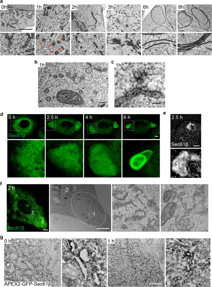
Fig. 3. ER whorls are formed from Sec61-containing tubular-vesicular precursors.
a Representative TEM micrographs of NRK cells treated with Tg for the indicated times. Regions of interest (outlined with white dashed lines) are magnified at the bottom. Red arrows in the lower 1 h panel indicate vesicles. Scale bar, 2 μm. b A TEM micrograph from a at 1 h. Scale bar, 200 nm. c A representative TEM micrograph of an NRK cell treated with Tg for 1 h. The block is processed to 300-nm-thick sections for observation. Scale bar, 100 nm. d NRK cells stably expressing GFP-Sec61β were treated with Tg and time-lapse images were acquired by Opera Phenix microscopy with 60× confocal mode. Regions of interest are magnified at the bottom. Scale bar, 5 μm. e NRK cells stably expressing GFP-Sec61β were treated with Tg for 2.5 h and visualized by GI-SIM at 100 nm resolution. Scale bar, 2 μm. The region of interest is magnified at the bottom. f CLEM imaging of a GFP-Sec61β-expressing NRK cell treated with Tg for 2 h. Left, the confocal image; middle, the TEM image; right, the enlarged images of the regions of interest outlined in the middle panel. 1, enlarged area of the GFP-Sec61β-condensed area; 2, enlarged area outside of the GFP-Sec61β-condensed area. Scale bar, 5 μm. g TEM image showing the DAB staining pattern in NRK cells stably expressing APEX2-GFP-Sec61β after treatment with Tg for 0 or 1 h. Scale bar, 2 μm.

