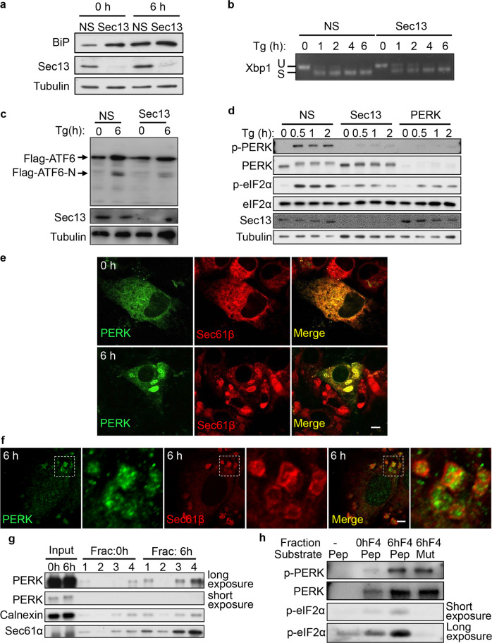
Fig. 6. COPII machinery is required for PERK activation.
a NRK cells were transfected with NS or Sec13 siRNA. Cells were treated with Tg for 0 or 6 h and analyzed by western blot using an antibody against BiP. b NRK cells were transfected with NS or Sec13 siRNA, and treated with Tg for the indicated times. Total RNA was subsequently extracted for semiquantitative RT-PCR analysis of XBP1 mRNA species (Xbp1 S: spliced XBP1 mRNA band; Xbp1 U: unspliced XBP1 mRNA band). c Flag-ATF6-expressing NRK cells were transfected with NS or Sec13 siRNA. Cells were treated with Tg for 0 or 6 h and analyzed by western blot using an antibody against Flag. d NRK cells were transfected with NS or Sec13 siRNA. Cells were treated with Tg for the indicated times and analyzed by western blot using an antibody against phospho-PERK (Thr980) and phospho-eIF2α (Ser51). e RFP-Sec61β-expressing NRK cells transfected with GFP-PERK were treated with Tg for 0 or 6 h and then observed by confocal microscopy. Scale bar, 5 μm. f Immunostaining of endogenous PERK and RFP in RFP-Sec61β-expressing NRK cells treated with Tg for 6 h. Scale bar, 5 μm. Regions outlined with white dashed lines are magnified. g NRK cells treated with Tg for 0 or 6 h were homogenized and the lysates were subjected to 1000× g centrifugation to discard the nuclei. The supernatant was ultracentrifuged in OptiPrep density gradient medium. The distribution of PERK in the fractions was monitored by western blot. 1 is the top fraction. h In vitro protein kinase assays. Membranes were collected from Fraction 4 of NRK cells treated with Tg for 6 h as described in g and mixed with ATP and the peptides eIF2α p(45–56) or mutant eIF2α p(45–56, S51A). Kinase reactions were resolved on SDS-PAGE and visualized by immunoblotting using an antibody against phospho-eIF2α (Ser51).

