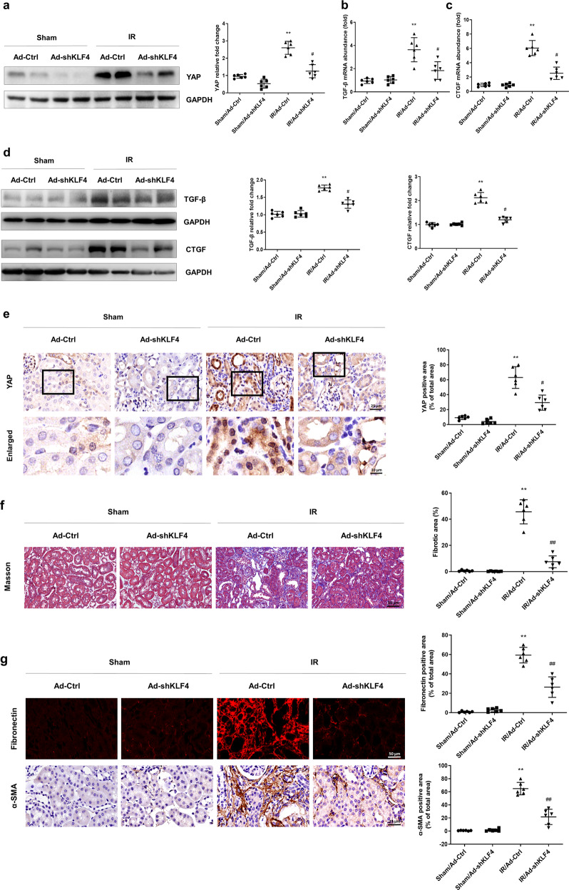
Fig. 3. KLF4 promotes progressive renal fibrosis after IR induction by upregulating YAP expression.
a Western blotting was conducted to detect the protein level of YAP in the four groups as indicated. The mRNA levels of TGF-β (b) and CTGF (c) were determined for the four groups as indicated. d Western blotting was conducted to determine the protein levels of TGF-β and CTGF in the four groups as indicated. e IHC was performed to visualize the distribution of YAP. Boxed areas are enlarged and presented in the figures below. Quantitative analyses of the YAP-positive area for the four groups as indicated. f Masson’s trichrome staining was used to detect collagen deposition in the four groups. Quantitative analyses of the extent of fibrosis. g Representative immunostaining images show fibronectin and α-SMA expression in the four groups. n = 6 mice in each group. Data are presented as the means ± SEM. **P < 0.01 versus the sham/Ad-Ctrl group. #P < 0.05, ##P < 0.01 versus the IR/Ad-Ctrl group, IR ischemia-reperfusion, IHC immunohistochemistry.

