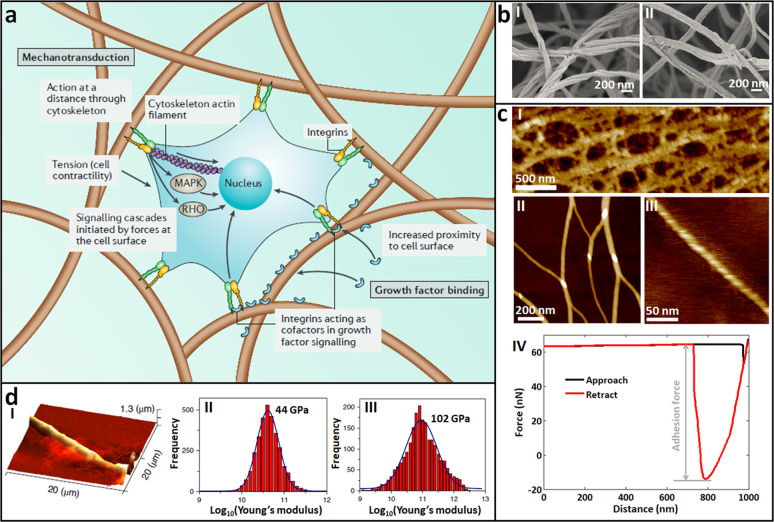
Fig. 11. AFM for visualizing the fine structures and measuring the mechanics of single ECM nanofibrils.
a Schematic illustrating the effect of fibrillar structures on mechanotransduction. Cells can exert forces on the fibers through cell-surface proteins and the cytoskeleton, which are both mechanically coupled to the ECM, initiating signaling pathways via mitogen-activated protein kinase (MAPK) and the RHO family of GTPases (RHO). Reprinted with permission from Ref. [156]. Copyright 2019 Springer Nature. b SEM images of collagen (I) and fibrin (II) gels, respectively. Reprinted with permission from Ref. [159]. Copyright 2015 Authors. c Imaging the fine structures of ECM and measuring the adhesion force of ECM by AFM [160, 161]. I Porous network structures of ECM visualized by large-size AFM scanning. II, III Single nanofibrils visualized by small-size AFM scanning. IV A typical force curve obtained on ECM for evaluating the adhesive capabilities of ECM. d Measuring the stiffness of single nanofibrils by AFM. I AFM image of tripeptide fibers. Statistical histogram of the Young’s modulus of the nanofibrils formed by Pro-Phe-Phe (II) or Hyp-Phe-Phe (III). Reprinted with permission from Ref. [164]. Copyright 2019 Springer Nature.

