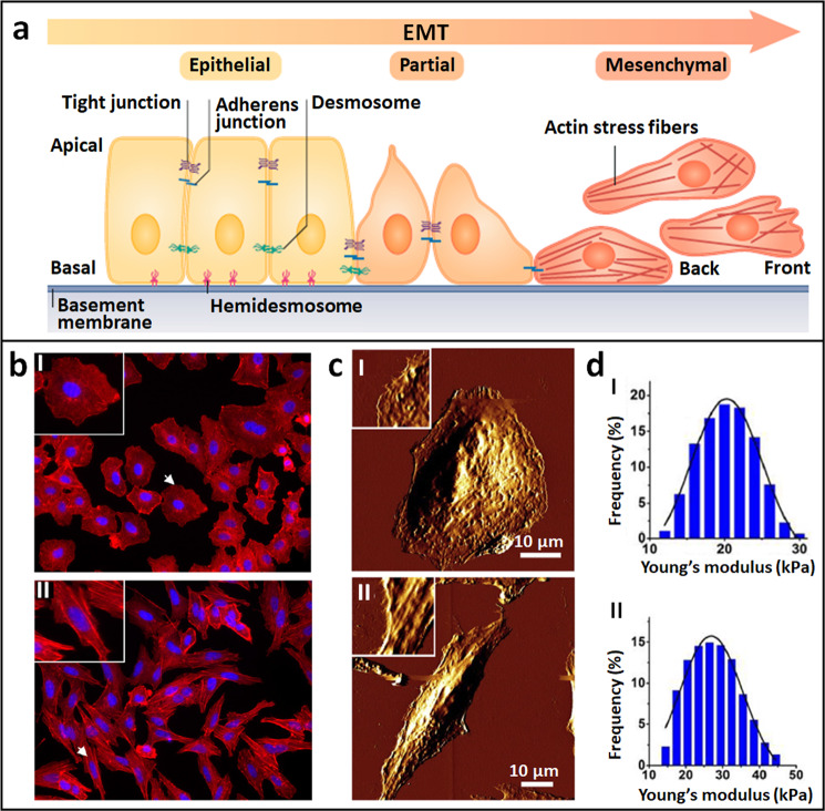
Fig. 4. Mechanical changes of cells in the process of EMT revealed by AFM.
a Schematic of EMT process. Epithelial cells displaying apical–basal polarity are held together by tight junction, adherens junctions, and desmosomes. Epithelial cells are tethered to the underlying basement membrane by hemidesmosomes. Induction of EMT results in cellular changes that include the disassembly of epithelial cell–cell junctions and the dissolution of apical–basal cell polarity. The loss of epithelial features is accompanied by acquisition of mesenchymal features. Mesenchymal cells display front-to-back polarity and have a reorganized cytoskeleton. After EMT, cells become motile and acquire invasive capabilities. Reprinted with permission from Ref. [58]. Copyright 2019 Springer Nature. b–d Cellular changes during EMT induced by TGF-β1. Cells before TGF-β1 treatment (I) and after TGF-β1 treatment (II). b Confocal fluorescent images. F-actins were stained with red fluorescein and nuclei were stained with blue fluorescein. The insets are the enlarged view of individual cells denoted by the arrows. c AFM morphological changes of cells. The insets are the enlarged view of local structures of the cells. d Statistical histograms of cellular Young’s modulus. b, c are reprinted with permission from Ref. [61]. Copyright 2012 Elsevier Inc. d is reprinted with permission from Ref. [62]. Copyright 2018 Elsevier B.V.

