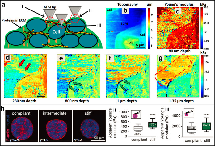
Fig. 7. Probing the mechanics of tumor cell spheroids by AFM.
a–g Visualizing the heterogeneous stiffness signature of tumor cell spheroids at different depths by analyzing the force curves obtained on spheroids. a Schematic showing that three types of nanomechanical topographies are identified during AFM indentation measurements, including collagen type I stress fibers (I), the interface of cell membrane and ECM (II) with high stiffness, and cells embedded deep inside the ECM (III). b Topography image and c–g corresponding stiffness images of a tumor spheroid at different depths. The double red arrows denote the collagen type I stress fibers, the double green arrows denote the interface of cell membrane and ECM, and the dotted red square denotes the cells embedded deep inside the ECM. Reprinted with permission from Ref. [100]. Copyright 2019 Springer Nature. h Mechanical dynamics of tumor spheroids and tumor cells regulated by the rigidity of microenvironment. I Confocal fluorescent images of the spheroids grown in hydrogels of varying stiffness. II AFM indentation assays on the whole tumor spheroids. III AFM measurements on individual tumor cells isolated from the tumor spheroids. Reprinted with permission from Ref. [102]. The insets in (II, III) show the schematic of AFM measurements on tumor spheroids or single tumor cells, respectively. Copyright 2019 WILEY-VCH Verlag GmbH & Co. KGaA, Weinheim.

