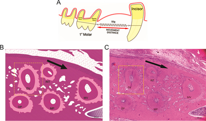
Figure 2.
Orthodontic tooth movement (OTM) (A) First left upper molar surrounded by a ligature wire and Niti spring and anchored in the upper incisor teeth. (B) Schematic model of the roots of the rat's left upper first molar. (C) Photomicrograph of the roots of the rat's left upper first molar. Five roots (MV, DV, INT, MP and DP) of the maxillary first molar; cross section. Dashed line highlights the DV root, chosen for histomorphometric evaluations. The arrow indicates the direction of the applied force. Bar indicates 500 µm, HE, ×40.

