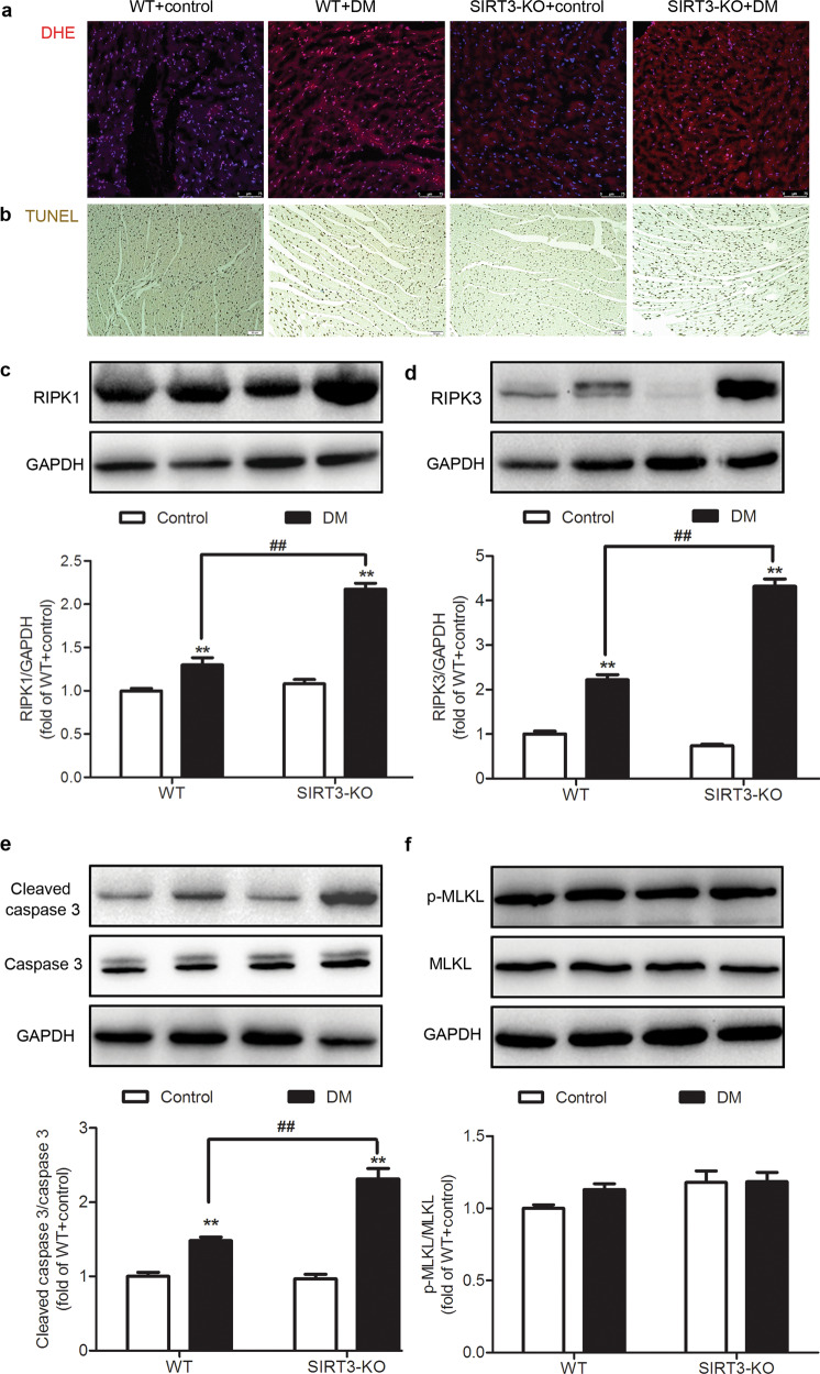
Fig. 4. SIRT3 deficiency promotes myocardial necroptosis in the diabetic mice.
After 12 weeks, the left ventricle of the myocardium was collected. a The level of superoxide anion in the myocardium was measured by DHE fluorescence probe. Bar = 75 μm. b The rate of apoptosis was assessed by TUNEL staining. Bar = 50 μm. c–f The protein expression levels of RIPK1, RIPK3, caspase 3, and MLKL in the myocardium were detected by Western blotting. Significance was determined by one-way ANOVA. Data are presented as the means ± SEM. **P < 0.01 vs the control group of the same genotype; ##P < 0.01 vs the DM group of WT mice, n = 6.

