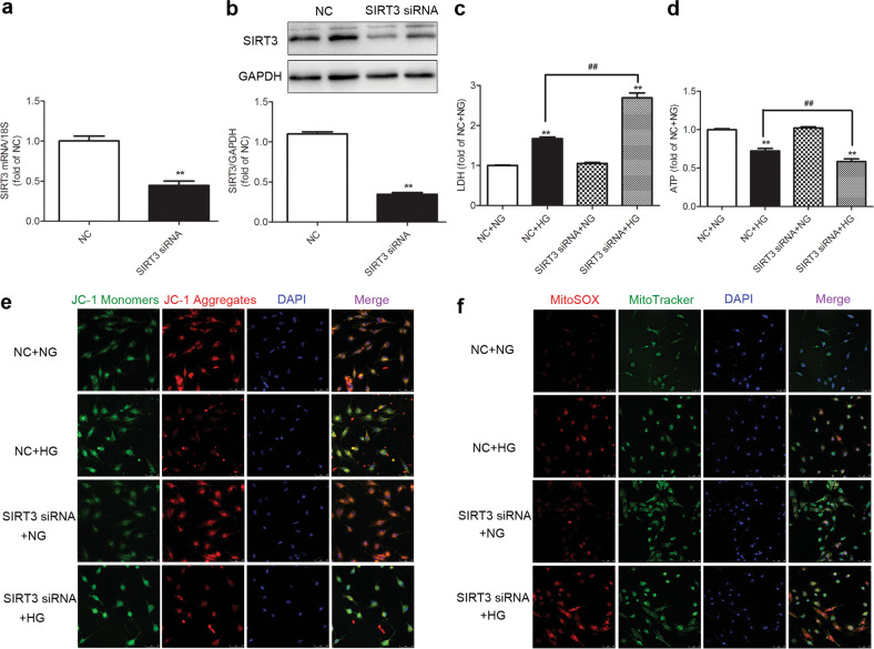
Fig. 5. SIRT3 silencing exacerbates cell injury and enhances oxidative stress in the high-glucose-stimulated cardiomyocytes.
SIRT3 siRNA and nonspecific control (NC) siRNA were transfected into cardiomyocytes. a, b After 48 h, SIRT3 mRNA and protein expression levels were measured by real-time PCR and Western blotting, respectively. **P < 0.01 vs the NC group, n = 6. c After 4 h, the cardiomyocytes were stimulated with normal glucose (5.5 mmol/L, NG) or high glucose (25.5 mmol/L, HG) for 48 h. LDH level in the medium was measured. d ATP levels in cardiomyocytes were measured. e The mitochondrial membrane potential (Δψm) of the cardiomyocytes was measured with JC-1 staining. Bar = 50 μm. f Mitochondrial superoxide was detected with MitoSOX. Mitochondrial localization of the MitoSOX signal was confirmed by MitoTracker green. Bar = 75 μm. Significance was determined by one-way ANOVA. Data are presented as the means ± SEM. **P < 0.01 vs the NG-treated cardiomyocytes transfected with the same siRNA; ##P < 0.01 vs the NC + HG-treated cardiomyocytes, n = 8.

