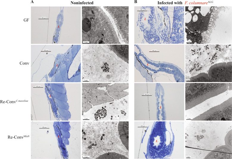
Fig. 5. Intestine of F. columnare infected germ-free zebrafish displays severe disorganization compared to conventional and reconventionalized larvae.
Germ-free, conventional and reconventionalized zebrafish larvae. Reconventionalized zebrafish were inoculated at 4 dpf with Mix9 or C. massiliae. a Representative picture of intestines of noninfected larvae. Fish were fixed for histology analysis or electron microscopy at 7 dpf. b Representative picture of intestines of infected larvae exposed at 7 dpf to F.columnareALG. In (a and b): Left column: Toluidine blue staining of Epon-embedded zebrafish larvae for Light microscopy. Right column: Transmission electron microscopy at 7 dpf (right). L = intestinal lumen.

