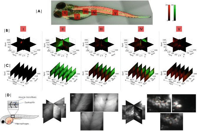
Figure 5.
Zebrafish embryo imaging. (A) Leica confocal images (tiled) of entire Zebrafish with a 10X objective lens. 48 h Zebrafish embryo with muscles and yolk stained with TRITC and BODIPY respectively show the distribution of lipids and muscles. (B) Reconstructed volume images of specific sections (marked I-V) of embryo are also shown. (C) 2D slice images from the reconstructed volume. (D) Images of muscle structure (planes, 26, 30 and 33) and primitive macrophages (planes, 12, 29 and 57) 24 hours post fertilization. (See, Supplementary 7 for details.

