Fig. 5.
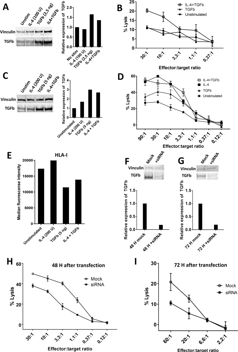
Recognition and killing of cancer cells by TGFβ-15-specific T cells depends on the expression of TGFβ by cancer cells. A Western blot (WB) analysis of intracellular expression of TGFβ in THP-1 cells that were stimulated for 48 h with either IL-4 (100 U/mL) or TGFβ (2.5 ng/mL) or both in combination. B Killing of THP-1 cells that were stimulated for 48 h with either TGFβ (2.5 ng/mL) or both with IL-4 (100 U/mL) and TGFβ (2.5 ng/mL). C Western blot analysis of intracellular expression of TGFβ in THP-1 cells that were stimulated for 48 h with either IL-4 (200 U/mL) or TGFβ (5 ng/mL) or both in combination. D Killing of THP-1 cells that were stimulated for 48 h with either IL-4 (200 U/mL) or TGFβ (5 ng/mL) or both in combination. E Expression of HLA-I in THP-1 cells treated with either IL-4 (200 U/mL) or TGFβ (5 ng/mL) or both in combination. F Western blot analysis of the expression of intracellular TGFβ in THP-1 cells after 48 h of transfection with TGFβ siRNA. G Western blot analysis of the expression of intracellular TGFβ in THP-1 cells after 72 h of transfection with TGFβ siRNA. H Cr51 release assay showing the killing of THP-1 cells mock-transfected or transfected with TGFβ siRNA at 48 H after transfection. I Cr51 release assay showing the killing of THP-1 cells mock-transfected or transfected with TGFβ siRNA at 72 H after transfection

