FIGURE 4.
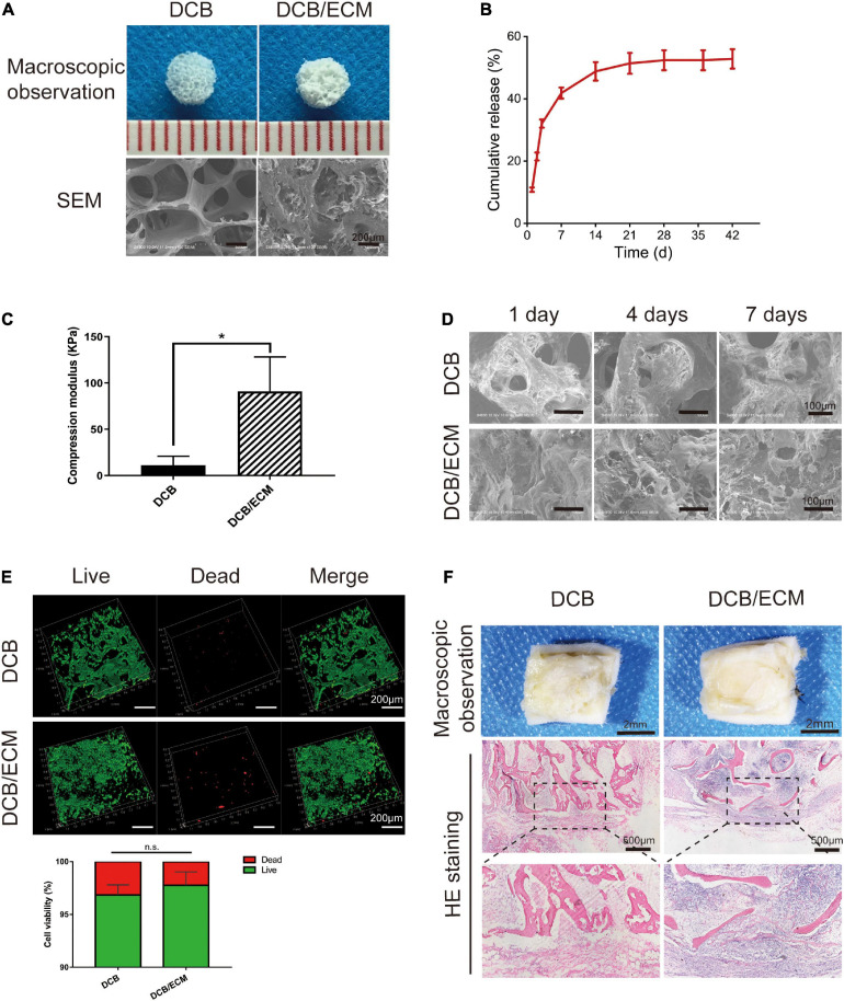
The physicochemical, biocompatibility, and immunogenicity properties of DCB and DCB/ECM scaffolds. (A) Macroscopic features and SEM of DCB and DCB/ECM scaffolds. (B) TGF-β3 release kinetics of the DCB/ECM/TGF-β3 scaffold. Values are presented as the means ± SDs (n = 3). (C) Compression modulus of DCB and DCB/ECM (scaffolds; values are presented as the means ± SD, n = 5). (D) SEM of DCB and DCB/ECM scaffolds on which IPFSCs were seeded for 1, 4, and 7 days. (E) Live/dead staining analysis of IPFSCs cultured in DCB and DCB/ECM scaffolds for 7 days. Representative 3D reconstruction images show live (green) cells and dead (red) cells. Values are presented as the means ± SDs (n = 3). (*p < 0.05, n.s. represents no significant difference). (F) Macroscopic observations and H&E staining of the immune responses of DCB and DCB/ECM scaffolds at 1-week postimplantation in rats.

