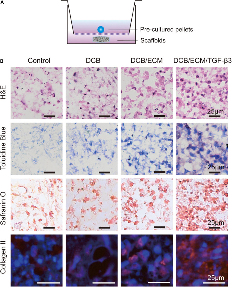
FIGURE 5.
Chondrogenic capacity of the different scaffolds in vitro. (A) Schematic illustrations of the coculture systems between pre-cultured pellets and the different scaffolds. (B) Histological and immunofluorescence analyses of chondrogenic pellets performed in a coculture system with different scaffolds. H&E, toluidine blue, safranin O, and collagen II immunofluorescence staining were used.

