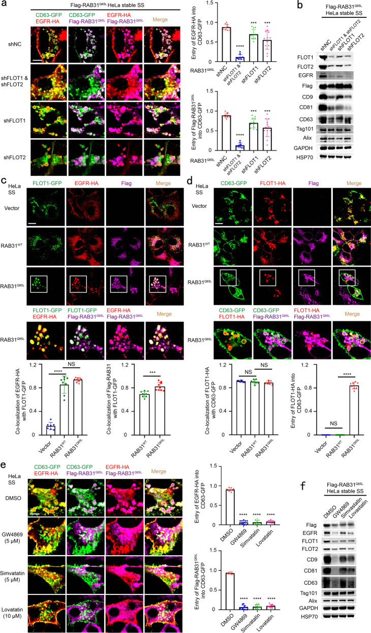
Fig. 3. Active RAB31 engages FLOTs to drive EGFR-containing ILV formation depending on cholesterol and ceramide in lipid raft microdomains.
a Left, immunofluorescence of EGFR-HA (red) and Flag-RAB31Q65L (magenta) with CD63-GFP (green) in Flag-RAB31Q65L stable HeLa cells stably expressing shNC (negative control), shFLOT1, shFLOT2 or shFLOT1 and shFLOT2 and transiently expressing EGFR-HA and CD63-GFP under serum starvation (SS). Right up panel, the ratio of entry of EGFR-HA into CD63-GFP-positive LE/MVE in shNC (n = 9 fields), shFLOT1 and shFLOT2 (n = 9 fields), shFLOT1 (n = 12 fields), shFLOT2 (n = 12 fields). Right low panel, the ratio of entry of Flag-RAB31Q65L into CD63-GFP-positive LE/MVE in shNC (n = 9 fields), shFLOT1 and shFLOT2 (n = 9 fields), shFLOT1 (n = 12 fields), shFLOT2 (n = 12 fields). b Western blotting analyses of the concentrated conditional media from the indicated stable HeLa cells used in a. c Up panels, immunofluorescence of EGFR-HA (red) and Flag-RAB31 (magenta) with FLOT1-GFP (green) in the indicated stable HeLa cells transiently expressing EGFR-HA and FLOT1-GFP under SS. Low panel left, the ratio of co-localization of EGFR-HA with FLOT1-GFP-positive vesicle in Vector (n = 7 fields), RAB31WT (n = 8 fields) and RAB31Q65L (n = 9 fields). Low panel right, the ratio of co-localization of Flag-RAB31 with FLOT1-GFP-positive vesicle in RAB31WT (n = 8 fields) and RAB31Q65L (n = 9 fields). d Up panels, immunofluorescence of FLOT1-HA (red) and Flag-RAB31 (magenta) with CD63-GFP (green) in the indicated stable HeLa cells transiently expressing FLOT1-HA and CD63-GFP under SS. Low panel left, the ratio of co-localization of FLOT1-HA with CD63-GFP-positive LE/MVE in Vector (n = 7 fields), RAB31WT (n = 7 fields) and RAB31Q65L (n = 8 fields). Low panel right, the ratio of entry of FLOT1-HA into CD63-GFP-positive LE/MVE in Vector (n = 7 fields), RAB31WT (n = 7 fields) and RAB31Q65L (n = 8 fields). e Left, immunofluorescence of EGFR-HA (red) and Flag-RAB31Q65L (magenta) with CD63-GFP (green) in Flag-RAB31Q65L stable HeLa cells transiently expressing EGFR-HA and CD63-GFP and treated with DMSO, 5 μM GW4869, 5 μM simvastatin or 10 μM lovastatin under SS. Right up panel, the ratio of entry of EGFR-HA into CD63-GFP-positive LE/MVE in DMSO (n = 8 fields), GW4869 (n = 11 fields), simvastatin (n = 13 fields) and lovastatin (n = 12 fields). Right low panel, the ratio of entry of Flag-RAB31Q65L into CD63-GFP-positive LE/MVE in DMSO (n = 8 fields), GW4869 (n = 11 fields), simvastatin (n = 13 fields) and lovastatin (n = 12 fields). f Western blotting analyses of the concentrated conditional media from the indicated stable HeLa cells used in e. All data are means ± SD. Unpaired t-test was used to analyze the difference between the two groups. ****P < 0.0001, ***P < 0.001, NS, no statistical significance. Scale bars, 10 μm.

