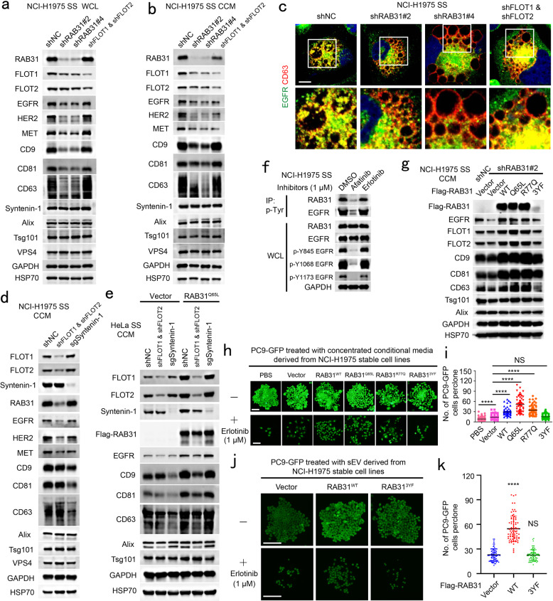
Fig. 6. EGFR phosphorylates RAB31 to drive EGFR into exosomes and the exosomes promoted by RAB31 mediate resistance to erlotinib.
a Western blotting analyses of whole-cell lysates (WCL) from the indicated NCI-H1975 cells stably expressing shNC, shRAB31 or shFLOT1 and shFLOT2. b Western blotting analyses of the concentrated conditional media from the indicated stable NCI-H1975 cells used in a under serum starvation (SS). c Immunofluorescence of endogenous EGFR (green) and CD63 (red) in the indicated stable NCI-H1975 cells used in a under SS. d Western blotting analyses of the concentrated conditional media from the indicated stable NCI-H1975 cells under serum starvation (SS). e Western blotting analyses of the concentrated conditional media from the indicated stable HeLa cells under serum starvation (SS). f Western blotting analyses of WCL and immunoprecipitates (IP) from NCI-H1975 cells treated with afatinib or erlotinib under SS. g Western blotting analyses of the concentrated conditional media from the indicated stable NCI-H1975 cells under SS. h Representative clone images of PC9-GFP cells treated with the concentrated conditional media derived from the indicated stable NCI-H1975 cells without or with erlotinib. i Quantification of the numbers of each clone for h. Data are means ± SD of cell numbers in each clone with PBS (n = 75), Vector (n = 82), RAB31WT (n = 62), RAB31Q65L (n = 62), RAB31R77Q (n = 65) or RAB313YF (n = 84). j Representative clone images of PC9-GFP cells treated with the pure small EV (sEV) derived from the indicated stable NCI-H1975 cells without or with erlotinib. k Quantification of the numbers of each clone for j. Data are means ± SD of cell numbers in each clone with Vector (n = 71), RAB31WT (n = 80) or RAB313YF (n = 80). Unpaired t-test was used to analyze the difference between the two groups. ****P < 0.0001, NS, no statistical significance. Scale bars, 10 μm (c) and 100 μm (h and j).

