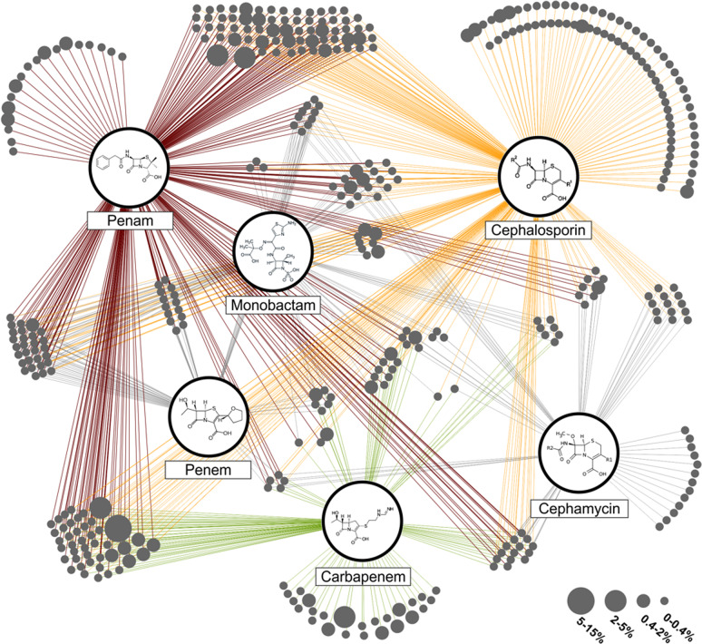
Fig. 5. Substrate spectrum of detected β-lactamases.
All assigned β-lactamases of the S. magellanicum from the Pirker Waldhochmoor, Austria, are displayed. The single β lactamases represented as bubbles (in dark grey) were grouped into clusters based on their reported substrate spectrum. Enzymes with the same substrate spectrum form one cluster. Connecting lines from the clusters to the β-lactam classes display the substrate specificity. Bubble size relates to the relative abundance of single enzymes within the whole β-lactamase pool. The three most abundant classes are penam (red), cephalosporin (orange) and carbapenem (green).

