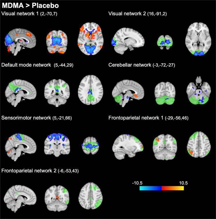
Fig. 1. Alterations in within-network functional connectivity (FC) after administration of MDMA compared with placebo (thresholded at p < 0.005, FWE, on the basis of a cluster-forming threshold of p < 0.001).
Resting-state networks identified in our data set are shown in green. MDMA significantly decreased FC within several networks (shown in blue). MDMA increased FC within parts of the frontoparietal networks (shown in red). After adjustment for potential confounds, alterations within the cerebellar network were no longer significant. These findings are nearly identical to alterations described after the administration of the hallucinogen LSD [4, 5]. The colorbar indicates the t values. X, Y, and Z values indicate MNI coordinates. Right is the right side of the brain.

