
FIGURE 3.
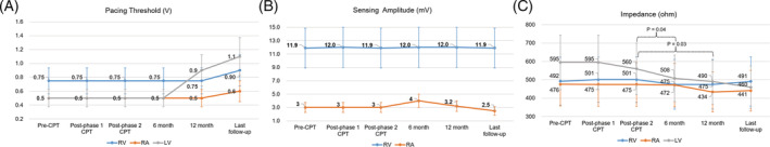
Lead integrity data according to follow‐up period after chest physiotherapy. Pacing threshold (A), sensing amplitude (B), and impedance (C) values are presented as median with interquartile range. P values were presented only when they were statistically significant. LV, left ventricular; RA, right atrial; RV, right ventricular