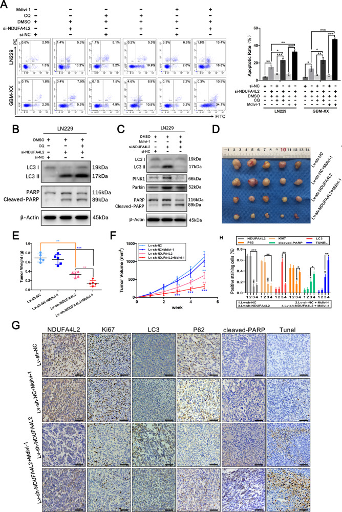
Fig. 3. NDUFA4L2 knockdown induces apoptosis, which is further enhanced by autophagy inhibitor treatment in vitro and vivo.
A Apoptosis rates in LN229 and GBM-XX cells treated with chloroquine (CQ; 10 μM) or Mdivi-1 (5 μM) were determined by flow cytometry (n = 3). B Protein levels of LC3, PARP, and cleaved-PARP in LN229 cells, following treatment with CQ (10 μM), were determined by western blotting. C Protein levels of LC3, PINK1, Parkin, PARP, and cleaved-PARP in LN229 cells, following treatment with Mdivi-1 (5 μM), were determined by western blotting. D–F Nude mice carrying tumors from LN229-transfected (sh-NDUFA4L2 or sh-NC by lentivirus) cells untreated or treated with Mdivi-1 (3 mg/kg) are shown (n = 5). The average tumor weight for each group was calculated. Tumor growth curves were calculated for each week. G-H NDUFA4L2, Ki67, LC3, p62, and cleaved-PARP expression levels, as well as positive cell numbers, were determined by immunohistochemical staining; apoptosis was assessed by TUNEL assay. Scale bar = 50 μm. Means ± SDs of triplicate experiments are plotted. *p < 0.05, **p < 0.01, ***p < 0.001.

