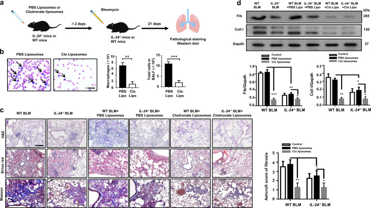
Fig. 4. Depletion of macrophages abolishes the protective effect conferred by IL-24 deficiency on BLM-induced lung injury and fibrosis.
a A schematic diagram for the macrophage depletion. Macrophages were depleted by intratracheal injection of clodronate liposomes, and injection of PBS liposomes were served as the controls. b Clodronate liposomes efficiently depleted macrophages in the lungs; macrophages were almost undetectable in the BALF of clodronate liposomes-treated mice along with a significant reduction in total cell numbers, Scale bar, 25 μm. Error bars represent means ± SEM (n = 5). Statistical analysis was performed using the Student’s t test (**p < 0.01; ***p < 0.001). c Depletion of macrophages restored IL-24−/− mice with manifestations similar as WT mice following BLM induction, as evidenced by the comparable histological changes and Ashcroft scores. Left panel: representative results for H&E, Sirius red, and Masson staining. Scale bar, 50 μm. Right panel: a bar graph showing the semiquantitative Ashcroft scores for the severity of fibrosis. Error bars represent means ± SEM (n = 5). Statistical analysis was performed using one-way ANOVA (*p < 0.05; **p < 0.01). d Macrophage-depleted WT and IL-24−/− mice manifested comparable levels of collagen I and fibronectin expression in the lung following BLM induction. Error bar represents the mean ± SEM of five mice analyzed. Statistical analysis was performed using one-way ANOVA (*p < 0.05; **p < 0.01; ***p < 0.001). BALF bronchoalveolar lavage fluid, BLM bleomycin, Clo Lipo clodronate liposomes, Coll I collagen I, Fib fibronectin, WT wild type.

