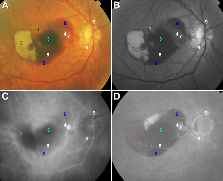
Fig. 1. Lesion components at baseline (October 2008), in aligned en face images.
Lesion boundaries are not shown for illustrative clarity. Components are numbered and color-coded for correspondence with Fig. 2. 1, sub-retinal pigment epithelium (RPE) fluid; 2, subretinal dehemoglobinized hemorrhage; 3, hemorrhage in Henle fiber layer; 4, sub-RPE hemorrhage and fluid; 5, subretinal hemorrhage; 6, sub-RPE hemorrhage; 7, aneurysm/polyp; 8, 9, peripapillary neovascularization. a Color fundus photograph; b red-free image; c mid phase indocyanine green angiogram; d late phase fundus fluorescein angiogram. Aneurysmal type 1 lesions 8-9 were best seen in a separately published late phase indocyanine green angiogram [26]; these are included here for completenes.

