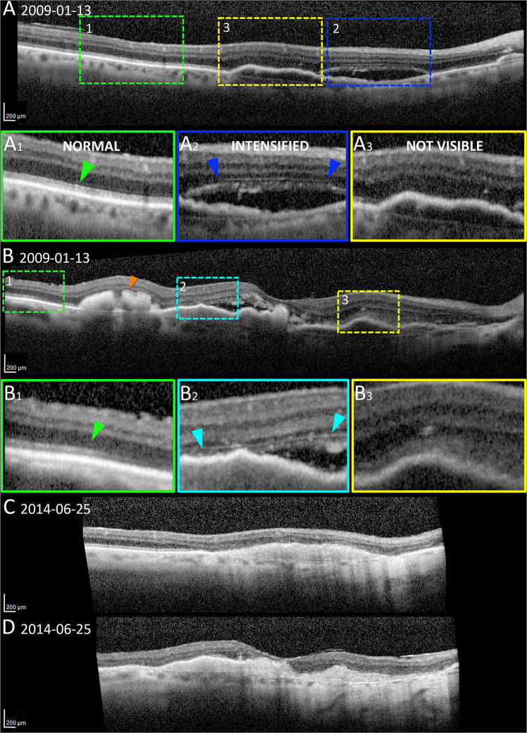
Fig. 2. Resolution of subretinal and intraretinal hemorrhage and exudation; variability of the external limiting membrane (ELM).
The ELM was seen as normal, condensed, and not visible on cross-sectional appearance on optical coherence tomography scans (a-b). a The subretinal exudation and trace amount of hemorrhage remained (blue frame) 3 months after the first photodynamic therapy (2009). The reflection of the associated ELM was intensified (magnified in A2, blue arrowhead) compared with the one visible in the temporal side (green frame, magnified in A1, green arrowhead). b Subretinal hyperreflectivity corresponding to the area of dehemoglobinized hemorrhage (orange arrowhead), and the hemorrhage in the HFL accompanied with intensification of ELM (teal frame, magnified in B2), were present. Areas with undetectable ELM (yellow frames, magnified in A3, B3) exhibited diffusely homogeneous hyperreflectivity that subsumed bands from RPE to HFL. c The follow-up scan of (a). The exudation and hemorrhage resolved, and the ELM was absent at the visit of 5 years later (2014). d The follow-up scan of (b). The hemorrhage resolved, and the ELM was absent.

