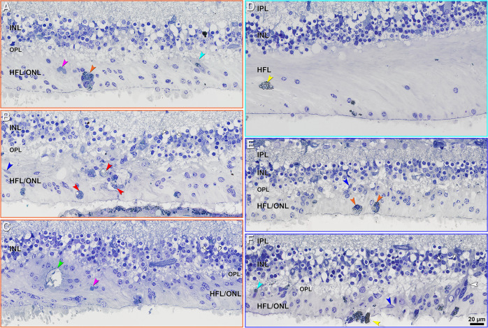
Fig. 4. Cells in Henle fiber layer (HFL)/outer nuclear layer (ONL) in areas with subretinal and intraretinal hemorrhage.
IPL inner plexiform layer; INL inner nuclear layer; OPL outer plexiform layer. Scale bar in (f) applies to all panels. All panels are color-coded in accordance with Fig. 1: orange frame (a–c) areas with dehemoglobinized subretinal hemorrhage; teal frame (d) areas with intraretinal hemorrhage; blue frame (e, f)- areas with non-dehemoglobinized subretinal hemorrhage. In areas with subretinal hemorrhage (a–c, e, f), the cellular changes are more common than in areas with intraretinal hemorrhage (d). In areas with intraretinal hemorrhage (d), the photoreceptor nuclei are mostly depleted, and the HFL is gliotic. Pink arrowheads, purple-stained debris; orange arrowheads, phagocytes with pigment granules; teal arrowheads, possible Müller cells with pigment granules; blue arrowheads, possible microglia; red arrowheads, phagocytes with lipids; green arrowhead, outer retinal tubulation; yellow arrowheads, migrated RPE cells; small white arrowheads, Müller cell bodies in INL; big white arrowheads, possible Müller cell bodies presenting in HFL/ONL with distortion of INL.

