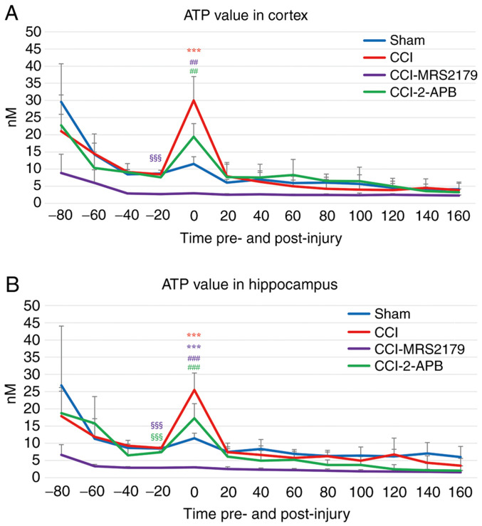
Figure 2.
ATP levels in the extracellular space in the (A) cortex and (B) hippocampus. The times shown for each 20-min dialysate represent the start of each sample collection relative to the time of injury. Low ATP baseline (-20 min pre-injury) levels were noted in the CCI-MRS2179 (cortex and hippocampus) and CCI-2-APB (hippocampus) groups compared with those of the sham injury controls. Large release of ATP was observed immediately after mild CCI injury in both the cortex and hippocampus. This increase was significantly attenuated by either MRS2179 or 2-APB treatment. The ATP values returned to the -20 min pre-injury levels at 20-40 min after injury. The color of each symbol denoting significant group differences correspond to the colors of the lines for each group shown in the legend. §§§P<0.001 vs. the sham group at -20 min pre-injury. ***P<0.001 vs. the sham group post-injury. ##P<0.01 and ###P<0.001 vs. the CCI group post-injury. 2-APB, 2-aminoethoxy diphenylborinate; CCI, controlled cortical impact; MRS2179, MRS2179 ammonium salt hydrate.

