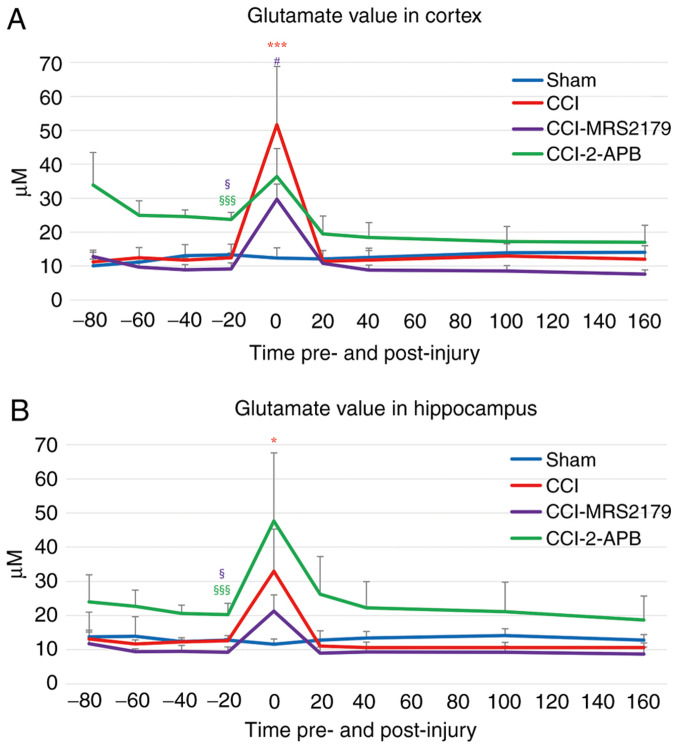
Figure 3.
Glutamate levels in the extracellular space in the (A) cortex and (B) hippocampus. The times shown for each 20-min dialysate represents the start of each sample collection relative to the time of injury. Low baseline (-20 min pre-injury) glutamate levels in the CCI-MRS2179 group and high baseline levels in the CCI-2-APB group compared with those of the sham controls were observed in both brain regions. A large release of glutamate was observed in the CCI group immediately after injury in both the cortex and hippocampus. This glutamate release was attenuated in the CCI-MRS2179 and CCI-2-APB groups. The color of each symbol denoting significant group differences correspond to the colors of the lines for each group shown in the legend. §P<0.05 and §§§P<0.001 vs. the sham group at -20 min pre-injury. *P<0.05 and ***P<0.001 vs. the sham group post-injury. #P<0.05 vs. the CCI group post-injury. 2-APB, 2-aminoethoxy diphenylborinate; CCI, controlled cortical impact; MRS2179, MRS2179 ammonium salt hydrate.

