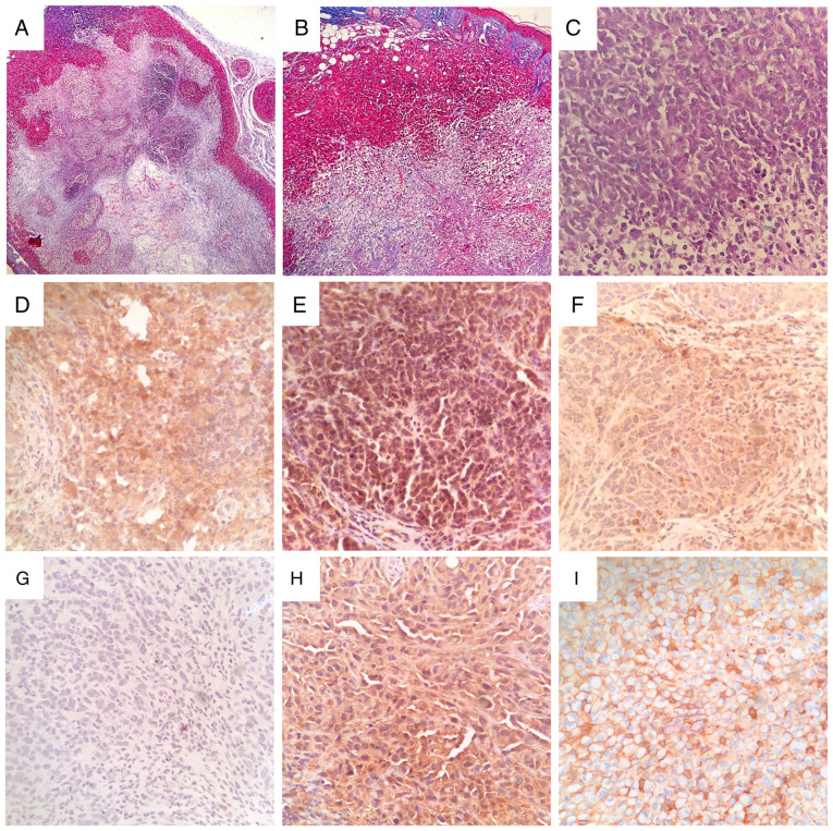
Figure 4.
Histopathological features and AVPR2 expression in MG-63 osteosarcoma xenografts. Representative images of tissue sections from rapidly growing MG-63 tumors stained using Masson's trichrome staining with aniline blue at x40 and x100 magnification (A and B, respectively) or hematoxylin and eosin at x400 magnification (C) AVPR2 presence in osteosarcoma tissue was detected using IHC on formalin-fixed paraffin-embedded MG-63 xenografts using primary antibodies against human AVPR2, polymer/HRP-liked secondary antibody and hematoxylin counterstaining. (D-F) Representative microphotographs of osteosarcoma xenografts after IHC staining. Magnification, x400. (G) Incubation with primary antibody was omitted. Human MDA-MB-231 (H) and MCF-7 (I) xenografts were used as positive controls for AVPR2 expression. Magnification, x400. AVPR2, vasopressin membrane receptor type 2; IHC, immunohistochemistry.

