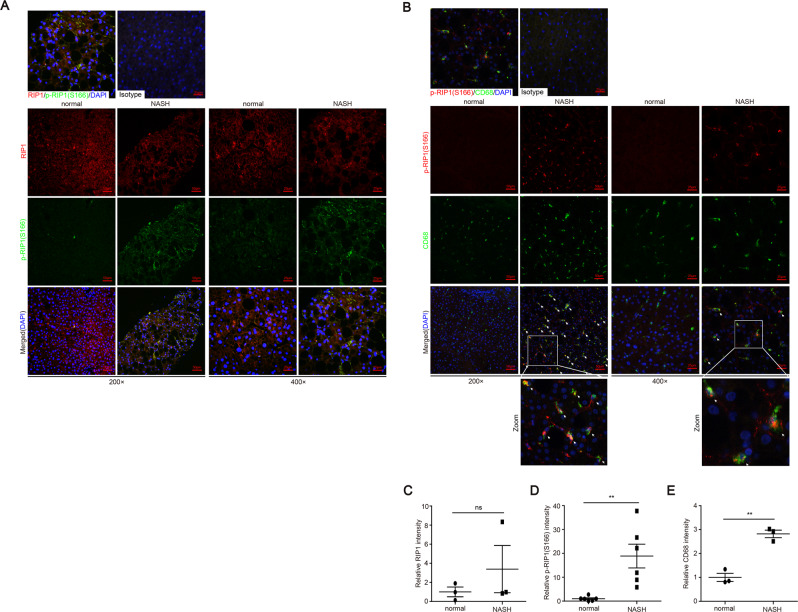
Fig. 6. RIP1 kinase was significantly activated in hepatic macrophages in human NASH.
A Representative images of liver tissue sections from control or NASH patients analyzed for the expression and phosphorylation of RIP1 at Ser166 by immunofluorescence staining. B Representative images of immunofluorescence double staining of phosphor-RIP1 (Ser166) (red) and CD68 (green). The nuclei were stained with DAPI (blue). C–E Quantitative analysis of RIP1-positive, phospho-RIP1-positive signals or CD68-positive signals in (A) or (B). Data are expressed as mean ± SEM. ns not significant, *p < 0.05, **p < 0.01, ***p < 0.001.

