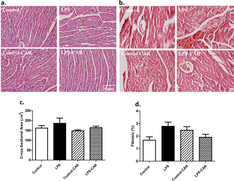
Fig. 2. Effect of CAR treatment (20 mg/kg, p.o.) on LPS challenge (4 mg/kg, i.p., for 6 h)-induced changes in cardiomyocyte cross-sectional area and interstitial fibrosis using H&E and Masson’s trichrome staining, respectively.
a Representative micrographs depicting H&E staining; b representative micrographs depicting Masson’s trichrome staining; c pooled data of cardiomyocyte cross-sectional area; and d pooled data of myocardial fibrosis; mean±SEM; n = 5 mice/group.

