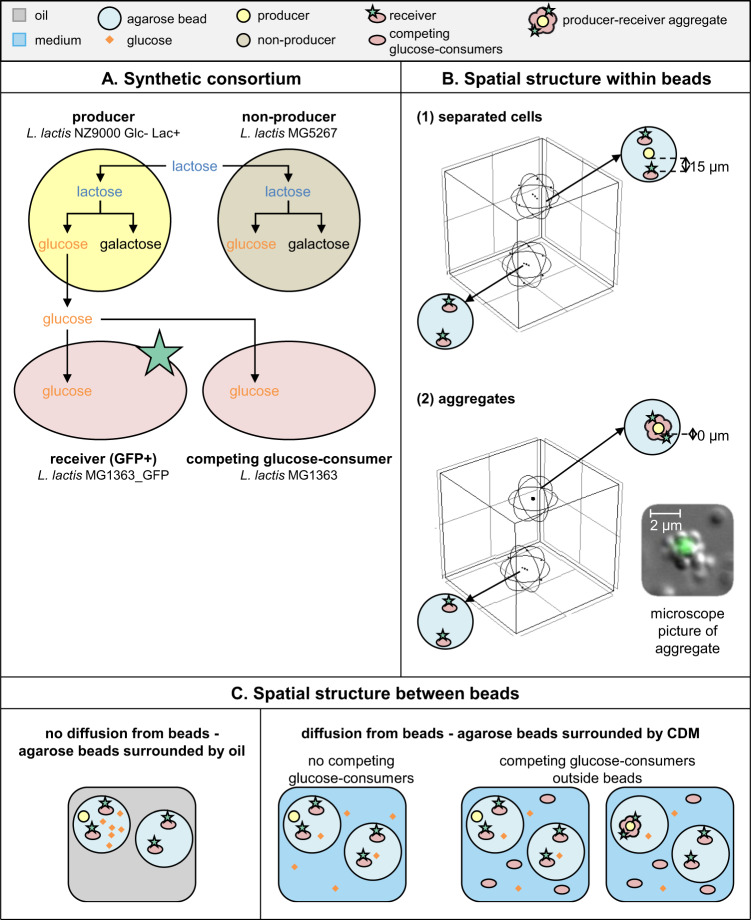
Fig. 2. Metabolic interactions in three-dimensional spatially structured environments.
A The four L. lactis strains that were used to make synthetic consortia: (1) “producers” which take up lactose and secrete glucose, (2) “receivers” which take up glucose and express GFP, (3) “non-producers” which take up lactose but do not secrete glucose and (4) “competing glucose-consumers” which take up glucose (Supplementary information section 2, Fig. S3). B The three-dimensional spatial structure within agarose beads. A distance of 15 µm between cells is comparable to a homogeneous distribution of 3 × 10⁸ bacteria/mL (Fig. S2). The microscopy image shows an aggregate where for visibility reasons a GFP-expressing cell is surrounded by nonfluorescent cells. Aggregates used in experiments were formed oppositely: a (non-)producer cell was surrounded by GFP-expressing receivers. C The three-dimensional spatial structure between agarose beads. Aggregates were only incubated in the presence of competing glucose-consumers.

