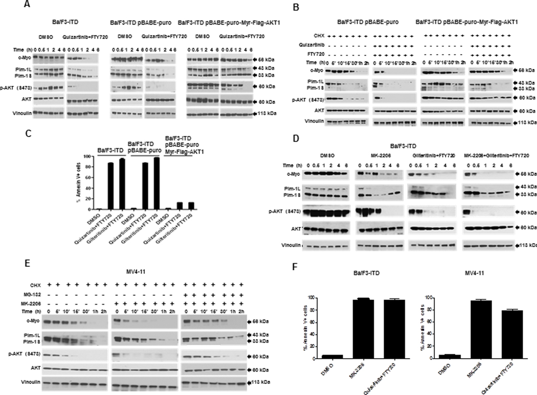
Figure 6. AKT inactivation is necessary and sufficient for c-Myc and Pim-1 downregulation and induction of apoptosis by PP2A-activating drug and FLT3 inhibitor co-treatment.

A. Constitutive AKT activation inhibits c-Myc and Pim-1 downregulation by combination treatment. c-Myc, Pim-1, p-AKT (S473) and AKT protein expression was measured by immunoblotting in parental Ba/F3-ITD cells and Ba/F3-ITD cells expressing myristoylated AKT (pBABE-Puro-Myr-FLAG-AKT1) or empty vector control (pBABE-Puro) treated with 1 nM quizartinib and 2 μM FTY720. B. Constitutive AKT activation inhibits increase in c-Myc and Pim-1 turnover in co-treated cells. c-Myc, Pim-1, p-AKT (S473) and AKT protein expression was measured by immunoblotting in Ba/F3-ITD cells expressing myristoylated AKT or empty vector control treated with 100 μg/ml cycloheximide (CHX) to block new protein translation, then with 1 nM quizartinib and 2 μM FTY720, or DMSO control. C. Constitutive AKT activation prevents induction of apoptosis by FLT3 inhibitor and PP2A activator combination. Apoptosis was measured by annexin V/PI labeling in parental Ba/F3-ITD cells and Ba/F3-ITD cells expressing myristoylated AKT or empty vector control treated with 1 nM quizartinib or 15 nM gilteritinib and 2 μM FTY720 for 48 hours. Constitutive AKT activation prevented induction of apoptosis (P<0.0005). D. AKT inhibition downregulates Pim-1 and c-Myc expression. c-Myc, Pim-1, p-AKT (S473) and AKT protein expression was measured by immunoblotting in Ba/F3-ITD cells treated with the AKT inhibitor MK-2206 (5 μM) and/or 15 nM gilteritinib and 2 μM FTY720. E AKT inhibition increases Pim-1 and c-Myc proteasomal degradation. c-Myc and Pim-1 expression was measured by immunoblotting in MV4-11 cells treated with 100 μg/ml cycloheximide (CHX), with and without the proteasome inhibitor MG-132 (20 μM), then with the AKT inhibitor MK-2206 (5 μM). F. AKT inhibition induces apoptosis of cells with FLT3-ITD. Apoptosis was measured in Ba/F3-ITD and MV4-11 cells treated with the AKT inhibitor MK-2206 (5 μM) or 1 nM quizartinib and 2 μM FTY720 or DMSO control for 48 hours. AKT inhibition induced apoptosis (P<0.0005).
