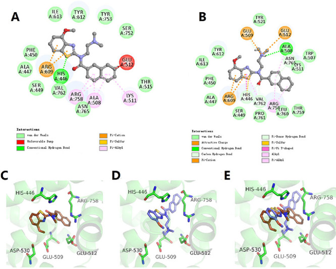
Figure 3.

The binding modes of 1r (A, C) and 1s (B, D) interacted with the antagonist-bound conformation of TRPC6 (PDB: 6uza). 1r and 1s were shown in brown and blue sticks in 3D mode. (E) Superimposed docking structures of TRPC6 in complex with 1s and 1r.
