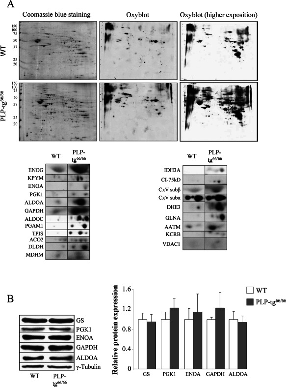
Figure 4.

Twenty one more markedly oxidized proteins identified in 6‐week‐old PLP‐tg66/66 mice in the spinal cord. A. Redox proteomics experiments were performed in 6‐week‐old PLP‐tg66/66 and their matched WT mice. Western blot with an anti‐ dinitrophenylhydrazine antibody at different exposure times to chemiluminescent reactive was performed to detect oxidized proteins (n = 3/genotype). B. All non‐mitochondrial oxidized proteins tested by Western blot in the spinal cord are normally expressed. Relative protein level expressed as a percentage of control and in reference to γ‐tubulin as a loading marker (n = 6/genotype). Values are expressed as the mean ± SD. Student's t‐test was used for statistical analysis; P < 0.05, **P < 0.01, ***P < 0.001.
