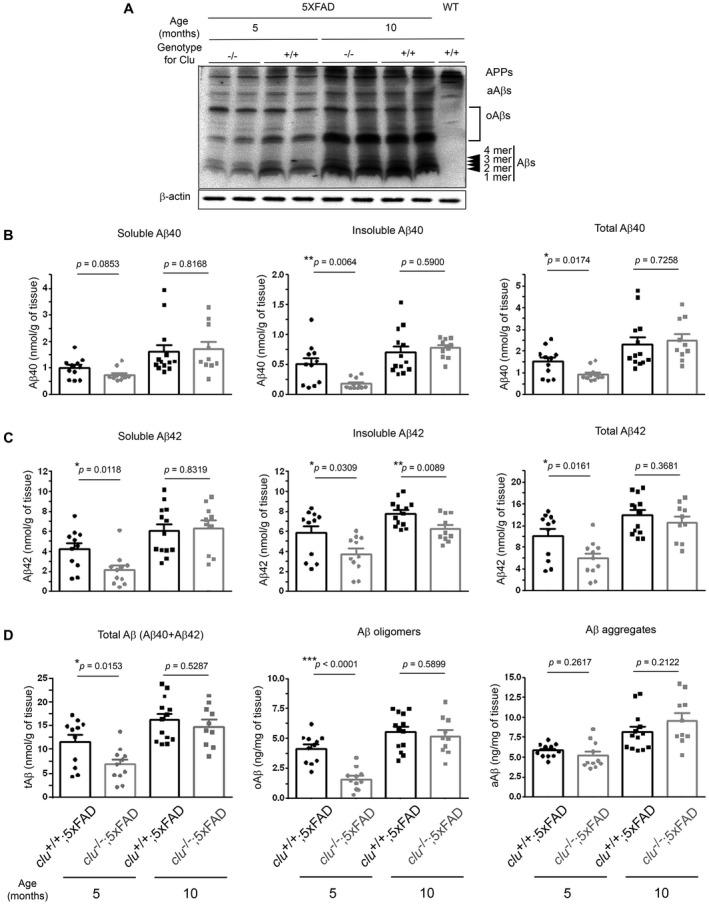
Figure 2.

Deposition of various pools of brain Aß in clu+/+;5×FAD and clu−/−;5×FAD mice. A. Representative Western blot shows expression or deposition of multiple sizes of Aß proteins in the brains of clu +/+;5×FAD and clu −/−;5×FAD mice at 5 or 10 months of age. After immunoblotting with anti‐Aß(1–16) antibody (6E10) (top panel), the blot was deprobed and reacted again with anti‐ß‐actin antibody as a loading control (bottom panel). Blot signals from a wild‐type littermate mouse are shown on the right end of the blot (lane 9). oAßs, Aß oligomers; aAßs, Aß aggregates. B–D. ELISA quantification shows the deposition of various pools of Aß proteins. Soluble (1st column) and insoluble (2nd column) Aß40 (B) or Aß42 (C) were analyzed from PBS‐ and SDS‐soluble fractions, and FA‐soluble fractions of brain lysates, respectively. Total levels of Aß40 (B, 3rd column), Aß42 (C, 3rd column) and Aß (D, 1st column) were calculated as the sum of soluble and insoluble Aß40, Aß42 and total Aß40 and total Aß42, respectively. Soluble Aß oligomers (oAß) were quantified from PBS‐ and SDS‐soluble fractions (D, 2nd column), whereas Aß aggregates (aAß) were analyzed from SDS extracts without ultracentrifugal fractionation (D, 3rd column). All ELISA analyses were performed in duplicate. Numbers (males and females) of animals included in each group were 11 (5 and 6; for 5‐month‐old clu +/+;5×FAD), 11 (5 and 6; for 5‐month‐old clu −/−;5×FAD), 13 (6 and 7; for 10‐month‐old clu +/+;5×FAD) or 10 (5 and 5; for 10‐month‐old clu −/−;5×FAD mice). *P < 0.05, **P < 0.01, or ***P < 0.001.
