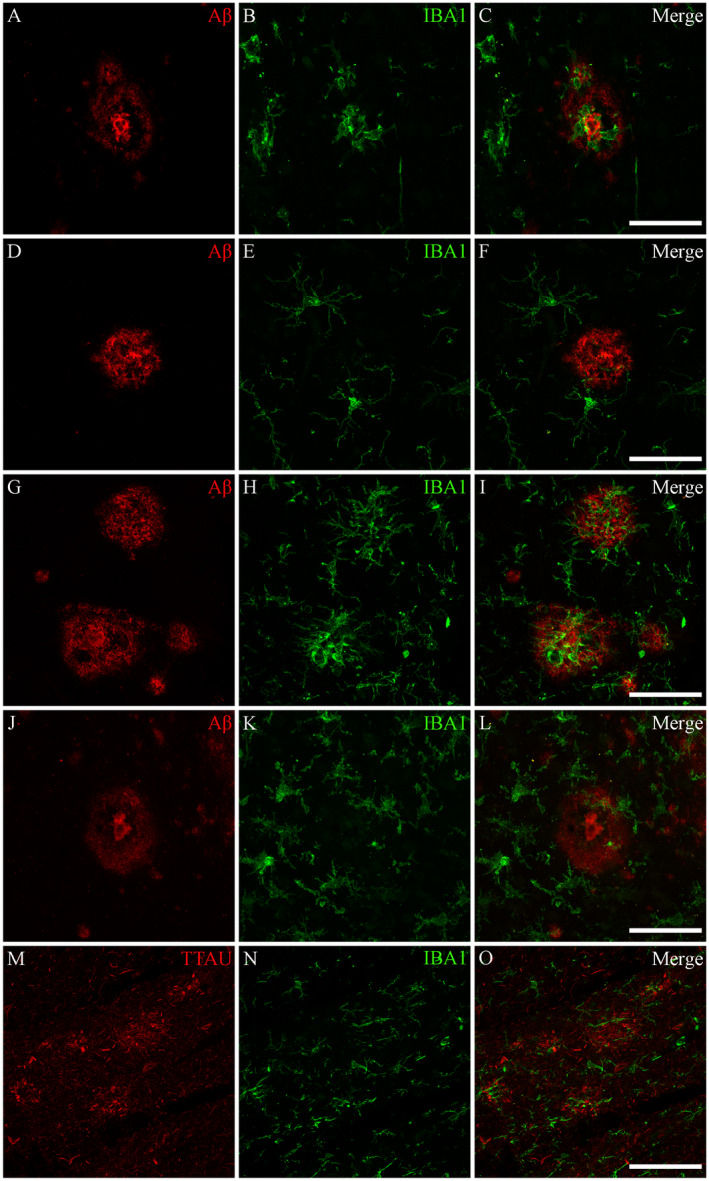
Figure 5.

Associations between microglia and AD plaques. Microglia were found to only occasionally cluster around tau‐positive plaques and Aβ‐immunopositive diffuse and cored plaques in both AD and control cases. Photomicrographs of immunofluorescent labeling for Aβ (A), IBA1 (B) and the merged image (C) from a control case with elevated AD‐type pathology (M23) demonstrates a cluster of ameboid microglia associated with a cored Aβ plaque. D–F. Ramified microglia and no microglial activation associated with a cored plaque of another control case (M19). Similarly, for AD cases, microglia were found to only occasionally cluster around Aβ plaques; as seen in the photomicrographs from an AD case (M09) demonstrating both significant clustering around diffuse and cored Aβ plaques (G–I), and an absence of microglial clustering (J–L). M–O. Tau‐positive plaques from the same AD case demonstrating an absence of microglial clustering. All photomicrographs were acquired using a Zeiss LSM 800 confocal microscope (40×/1.3 Oil). Scale bars = 50 μm.
