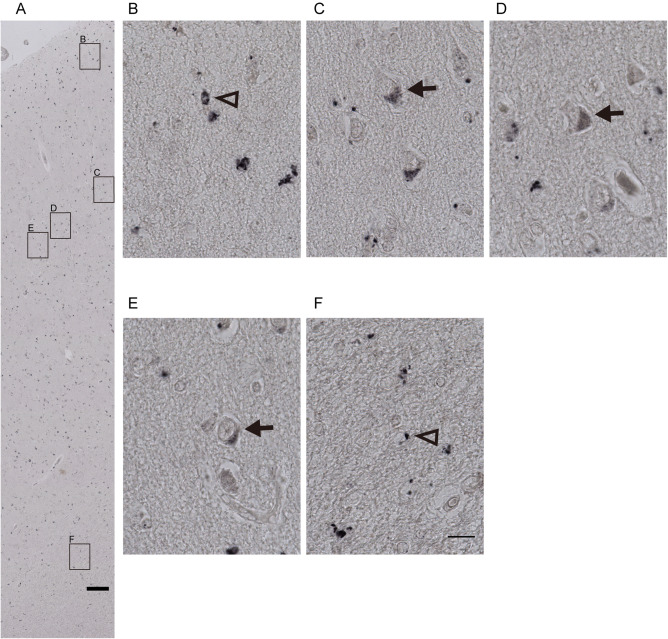
Figure 3.

Representative microphotographs of SULT2A1 IHC in the human anterior cingulate cortex. Immunohistochemical staining in the human anterior cingulate cortex (ACC) showed that SULT2A1 protein was localized from the pial layer to the white matter in ACC, A, and was present in both, nucleolus containing larger neurons (arrows in C–E in the grey matter) and in smaller glia‐cells (triangle in B in the non‐neuron containing layer 1 and F in the white matter). Scale bars: A = 200 µm; B–F = 25 µm.
