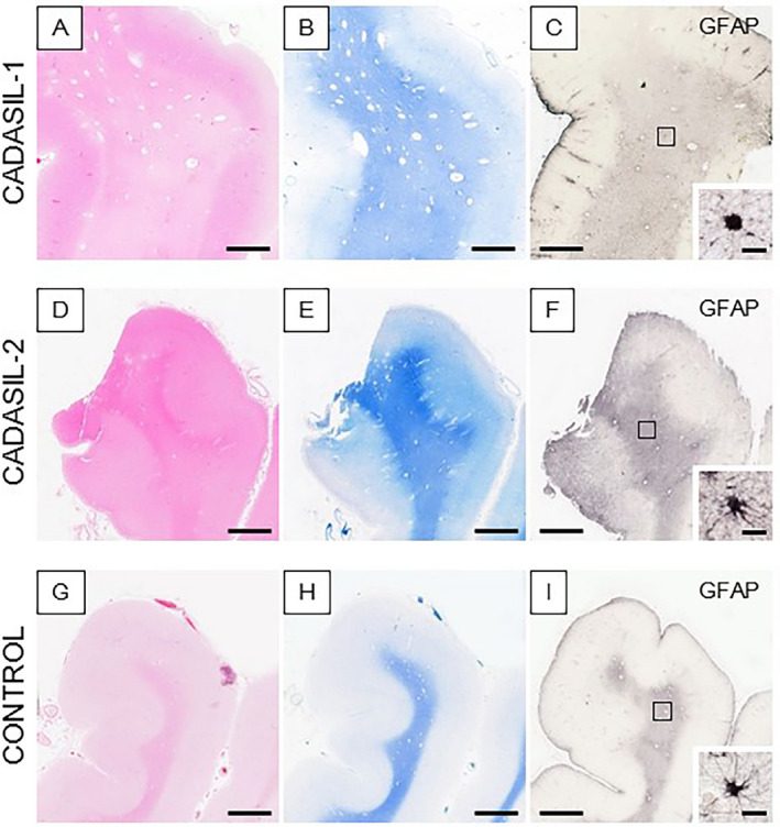
Figure 1.

Histological features in the anterior temporal pole of CADASIL and control subjects. (A–I) Images show H&E (A, D and G), LFB (B, E and H), and GFAP (C, F and I) stained sections in two CADASIL patients with NOTCH3 mutations (R133C and R558C) (A–F) and a similar age control (G–I). Insets in C, F, and I show profiles of single GFAP immunostained astrocytes; abnormal astrocytes (C and F); and normal appearing astrocyte (I). Consistent with MRI, the WM in both CADASIL patients also revealed numerous perivascular spaces. Magnification bar = 3mm (A–I) and in insets C, F, and I = 20μm.
