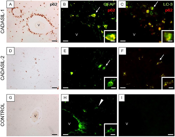
Figure 5.

Co‐localization of LC3, p62 and GFAP immunoreactivities in the WM of CADASIL subjects. (A–I) Sections from the anterior temporal lobe from a 61‐year‐old male with CADASIL carrying the R133C mutation (A–C), 55‐year‐old male with CADASIL carrying the R558C mutation (D–F) and age matched control cases (G–I) were immunostained for p62 (red) and GFAP (green) or LC3 (green). (A, D and G) Perivascular immunoreactivity of p62 was observed around >90% of the vessel profiles in CADASIL with R133C mutation (A), whereas less perivascular p62 immunoreactivity was observed in CADASIL with R133C mutation (D) and no apparent p62 immunoreactivity was observed in control (G). (B, C, E, F, H, and I) Compared to control (H and I) the merged immunofluorescent images from CADASIL subjects show strong co‐immunolocalization of p62 antigen in degenerating GFAP‐labeled astrocytes (arrows in B and E) which are also immunopositive for LC3 (arrows in C and F). (H–I) Normal astrocytes stained for GFAP with no apparent immunopositivity to p62 (H) (arrow head) and negative immunoreactivitiy for LC3 (I). Magnification bar = 50 μm (A, D and G), 25 μm (B, C, E, F and H) and in insets B, C, E, F and H =10μm. ‘v’ indicates a blood vessel (B, C, G, H and I).
