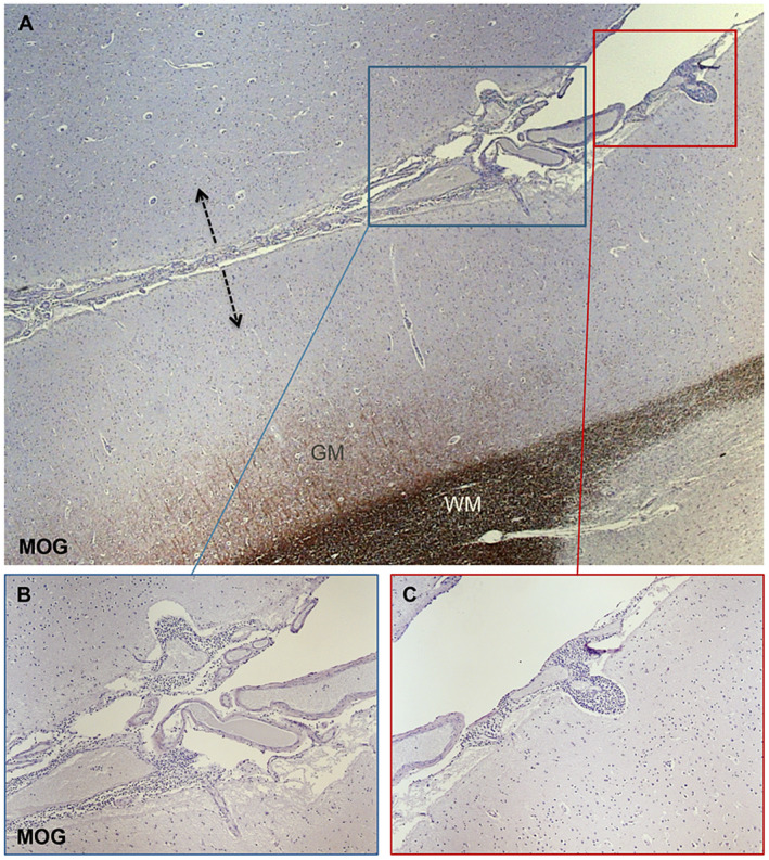
Figure 1.

Neuropathology assessment of subpial cortical demyelination associated with meningeal inflammatory infiltrates. MOG immunostaining (A‐C) allows the detection of subpial cortical lesions that are often expanding from both sides (arrows in A) of cerebral sulci as specular images. Within the meninges lining on the pial surface numerous extravasating inflammatory infiltrates are evident (B and C). Some of them appeared invading the adjacent, superficial cortical layers (B and C).
