Figure 1.

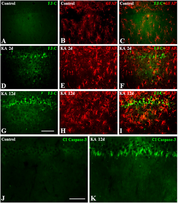
A–I. Photomicrographs depicting Fluoro‐Jade C labeled neurons (A, D, G), GFAP‐labeled astrocytes (B, E, H) and their spatial co‐distribution (C, F, I) in the hippocampus of control (A–C), 2day (D–F) and 12day (G–I) kainic acid‐treated rats. Note the lack of Fluoro‐Jade C labeling and proliferation of astrocytes in control rat hippocampus (A–C), whereas the loss of neurons and proliferation of astrocytes increased gradually in the hippocampus of kainic acid‐treated rats over 12day post‐treatment period. J and K, Photomicrographs depicting cleaved‐caspase‐3 labeling in the hippocampus of control (J) and 12day kainic acid‐treated (K) rats. Note the cleaved‐caspase‐3 labeled neurons following administration of kainic acid but not in control rat. Scale bar, 100 µM.
