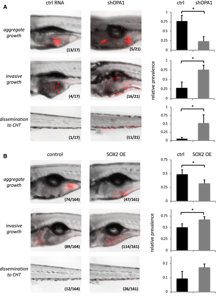
Figure 3.

A. OPA1 knock‐down increases invasiveness of LN319 cells in vivo. Left: Confocal pictures of zebrafish embryos effectively xenotransplanted with LN319 control cells (shRNA non‐coding, left) or LN319 shOPA1 knock‐down cells (right). LN319 cells (Cell Tracker, red) either established stable cell masses near the site of injection (aggregate growth, top), dispersed throughout the yolk sack (invasive growth, center) or disseminated to the caudal hematopoietic tissue (CHT) of the tail fin (bottom). Inlays indicate absolute animal numbers per condition. Right: Relative quantification of experimental outcomes documenting aggravated invasiveness and dissemination of OPA1 knock‐down vs. respective control cells. B. SOX2 overexpression (OE) increases invasiveness of LN319 cells in vivo. Experimental setup as above, except that TetON (control, left) and TetON mCherry‐SOX2 (SOX2 OE, right) cells had been pretreated with doxycycline (1 µg/ml for 24 h) to selectively induce SOX2 protein formation prior to transplantation. Right: Assay quantification verifying increased invasiveness and dissemination to CHT (by trend) also for SOX2 OE cells when compared to equally treated controls. Significance cut‐off‐: *P < 0.05.
