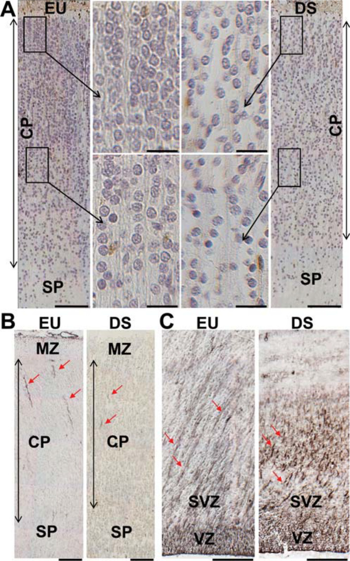
Figure 2.

Cytoarchitecture of the fusiform gyrus and inferior temporal gyrus in DS and euploid fetuses. A. Sections immunostained for calretinin and counterstained with hematoxylin from the inferior temporal gyrus of a euploid fetus (image on the left) and a DS fetus (image on the right) at GW21. Images in the middle are higher magnification of the boxed areas. Note the radial organization of the cortical plate of the euploid fetus and the more disorganized appearance and hypocellularity of the cortical plate of the DS fetus. Calibrations = 100 µm (low magnification images); 20 µm (high magnification images). B,C. Sections immunostained for vimentin from the cortical plate (B) and ventricular region of the inferior temporal gyrus (C) at GW21. The red arrows indicate processes of radial glia cells. Note the ordered organization of radial glia processes in the ventricular zone/subventricular zone of the euploid fetus. Calibrations = 100 µm. Abbreviations: CP, cortical plate; EU, euploid; DS, Down syndrome; MZ, marginal zone: SP, subplate; SVZ, subventricular zone; VZ, ventricular zone.
