
Figure 2.
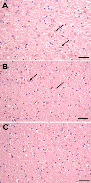
Temporal white matter from a patient with Type II AxD. A. The periventricular white matter is pale and contains RFs (arrows) and enlarged astrocytes. B. The deep white matter shows scattered RFs (arrows), but the degree of myelination appears normal. C. The astrocytes and the degree of myelination in U fibers appears normal. All Figures H&E. Scale Bars (A, B, C) 200 μm.