
Figure 3.
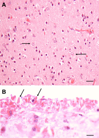
Isocortex of a Type I AxD patient. A. Isocortex does not show RFs, although some astrocytes are enlarged (arrows). B. RFs (2 marked by arrows) accumulate at the pial surface (top of image) in the endfeet of astrocytes. All Figures H&E. Scale Bars (A) 50 μm, (B) 150 μm.