
Figure 3.
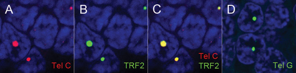
The large, ultra‐bight foci are specific for telomeric DNA. A. Telomere DNA is labeled with a Cy3‐labeled telomere sequence‐specific probe complementary to the G‐rich telomere repeat strand sequence (Tel‐C, red); B. Same area as in A, showing anti‐TRF‐2 antibody staining (green) (B); and C. co‐localization of A&B (yellow). D. FAM labeled telomere DNA probe (Tel‐G, green; guanine‐rich probe complementary to the C‐rich strand telomere repeat sequence) demonstrates a similar staining pattern, thus indicating the large, ultra‐bright foci consist of telomere DNA, which is G‐rich and not telomeric RNA, which is transcribed from only one strand of telomeric DNA, producing RNA consisting of the C‐rich telomere repeat sequence. For all images, DNA is stained with DAPI (blue).