
Figure 4.
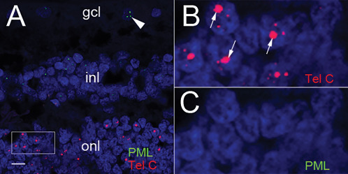
PML does not co‐localize with the large, ultra‐bright telomere DNA foci. A. Telomere DNA is labeled with a Cy3‐labeled telomere sequence‐specific probe complementary to the G‐rich telomere repeat sequence (Tel‐C, red) and PML nuclear bodies are labeled with an anti‐PML antibody and detected with a Cy5‐conjugated secondary antibody (green). PML nuclear bodies (green) are observed only in the retinal ganglion cell layer (arrowhead) and neurons of in the inner nuclear layer; however, not present in the rod photoreceptors containing large telomeric foci (red), scale: 10 µm. B. Higher magnification of box selected region in panel A highlighting the Cy3‐labeled telomeres (red) and large telomeric foci (arrows). C. Higher magnification of box selected region panel A, omitting telomere signals, showing no PML nuclear bodies in focus‐positive rod photoreceptor cells (panel B) and no PML nuclear bodies in any photoreceptor cells. For all images, DNA is stained with DAPI (blue).