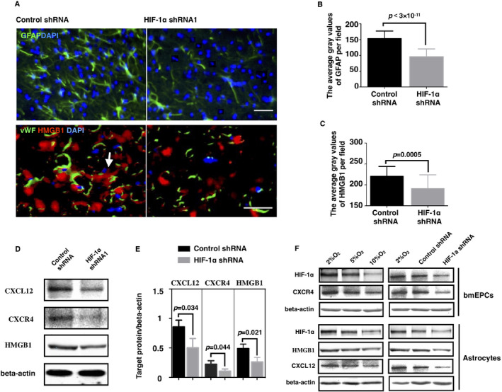
Figure 6.

Knockdown of HIF‐1α impaired the microenvironment that recruited bmEPCs. A. On the 7th day of MCAO, the brains were removed, and 20‐μm brain sections were prepared and fixed for immunostaining. The average gray value of the AOIs was used to quantify the HMGB1 expression and GFAP expression. B and C are the statistical analyses of panel (A) (n = 3). D, E Brains were isolated for western blotting on the 7th day after the MCAO surgery. F. Primary bone marrow‐derived EPCs and astrocytes were cultured under a gradient oxygen situation for 8 h. Then, the cells were harvested for the protein expression analysis of CXCR4, HMGB1, and CXCL12 by western blotting. Data were generated from at least 3 independent experiments. (n = 23–28 fields from 5 to 6 brains per group). Bar, 60 μm. Data are presented as the mean ± S.D.
