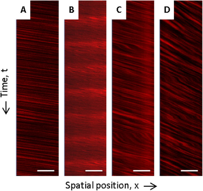
Figure 1.

Intravital RBC flow velocity measures using 2‐photon microscopy. A shows an exemplary line scan of an arteriole without any HA phenomena displaying unhampered RBC flow velocity. For comparison, B—D demonstrate line scans of arterioles characterized by RBC flow velocity disturbances. B thereby shows a line scan of an exemplary arteriole with luminal Dextran accumulations and C and D display line scans of vessels filled with non‐occlusive erythrocyte thrombi. HA, hypertensive arteriopathy; RBC, red blood cell. Scale bars = 20 μm; x‐axis—spatial position of RBCs; y‐axis—time t (10.000 ms from top to bottom of the line scan).
